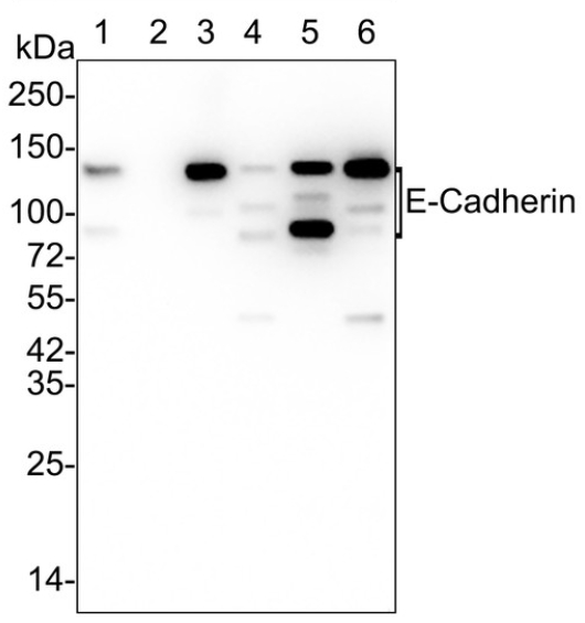
Anti-E-Cadherin antibody
Specifications
| Product Cat#: | AB-06-1475 |
| Product type: | Primary antibody |
| Antigen: | E Cadherin |
| Immunogen: | Recombinant protein |
| Species immunized: | Rabbit |
| Isotype: | IgG |
| Applications: | Western Blot ( 1:1000-1:5000 ); Immunohistochemistry ( 1:80-1:200 ); Immunocytochemistry ( 1:80-1:200 ); Immunoprecipitation; Immunofluorescence; Flow Cytometry ( 1:40-1:100 ) |
| Reactivity: | Human |
| Clonality (clone number): | Monoclonal ( SY0287 ) |
| Form: | Liquid |
| Buffer: | Tris-HCl buffer ( pH7.4 ), 1 % BSA, 40 % glycerol, 0.05 % NaN3. |
| Concentration: | 1 mg/ml |
| Purity: | Protein A affinity purified |
| Storage: | Aliquot and freeze at -20℃. Avoid multiple freeze/thaw cycles. |
| Alternative names: | Arc 1 Antibody |
| CADH1_HUMAN Antibody | |
| Cadherin 1 Antibody | |
| Cadherin 1 Type 1 E-cadherinAntibody | |
| Cadherin1 Antibody | |
| CAM 120/80 Antibody | |
| CD 324 Antibody | |
| CD324 Antibody | |
| CD324 Antigen Antibody | |
| Cdh1 Antibody | |
| CDHE Antibody | |
| E-Cad/CTF3 Antibody | |
| E-cadherin Antibody | |
| ECAD Antibody | |
| Epithelial cadherin Antibody | |
| Epithelial Calcium Dependant Adhesion Protein Antibody | |
| LCAM Antibody | |
| Liver Cell Adhesion Molecule Antibody | |
| UVO Antibody | |
| Uvomorulin Antibody |
Show More